Lietuvos chirurgija ISSN 1392–0995 eISSN 1648–9942
2023, vol. 22(4), pp. 249–252 DOI: https://doi.org/10.15388/LietChirur.2023.22(4).8
Pediatric Patient with Omental Cyst: A Rare Case (CT scan is not Enough to Differentiate from Other Abdominal Cysts)
Donny Aditia
Pediatric Surgery Division, Department of Surgery, Faculty of Medicine, Public Health, and Nursing, Universitas Gadjah Mada, Yogyakarta, Indonesia
E-mail: donnyaditiadr@mail.ugm.ac.id
Andi Dwihantoro
Pediatric Surgery Division, Department of Surgery, Faculty of Medicine, Public Health, and Nursing, Universitas Gadjah Mada, Yogyakarta, Indonesia
E-mail: andi.dwhivantoro@yahoo.com
Abstract. Background. Omental cysts are rare intra-abdominal tumors in children. It is challenging to diagnose since other big abdominal cysts are difficult to distinguish from in clinical and CT imaging. Aim. This case report aims to accurately and thoroughly determine the diagnosis based on the anamnesis, clinical and imaging examinations to choose the best treatment for the patient. Case Report. A 4-year-old girl reported having an enlarged belly in the past two months; the anamnesis provided the diagnosis. A mobile, 15 cm by 15 cm mass with dull percussion was discovered during clinical investigations in the top border. It was determined by anamnesis, physical examination, and imaging that the diagnosis was an ovarian cyst. Instead of an ovarian cyst, we found an omental cyst during the operation, thus we had to conduct an excision and omentectomy and postoperatively achieved favorable results. She was discharged three day after. Discussion. An abdominal CT scan showed an ovarian cyst. In laparotomy, we found a giant omental cyst, not originating from the ovaries, and did an omentectomy to excision the cyst. CT scan shows that the giant abdominal cyst is difficult to distinguish between omental, mesenteric, and ovarian cysts. Precise identification of stomach cysts is necessary. Also, doctors need to read the CT scan image more carefully. Conclusion. Giant omentum cysts are difficult to distinguish clinically and imaging from ovarian and mesenteric cysts. There is a need for a more detailed history, physical examination, and support, as well as a more thorough reading of the CT scan.
Keywords: abdominal cyst, CT scan, omental cyst, ovarium cyst, omentectomy, mesenteric cyst, giant abdomen cyst.
Received: 2023/08/10. Accepted: 2023/10/09.
Copyright © 2023 Donny Aditia, Andi Dwihantoro. Published by Vilnius University Press. This is an Open Access article distributed under the terms of the Creative Commons Attribution Licence, which permits unrestricted use, distribution, and reproduction in any medium, provided the original author and source are credited.
Background
Omental cyst are rare intra-abdominal tumors that mostly affect children. They frequently have symptoms that are similar to those of other, more prevalent abdominal illnesses and usually are unintentionally found on imaging. Omental cysts are rare intra-abdominal tumors in children. Rarity of large omental cysts, we therefore report a case of a large omental cyst in a 4-year-old girl who presented with an enlarged abdomen and what we initially mistook for an ovarian cyst from the abdomen CT scan, it’s often difficult to identify the origin of intra-abdominal tumors as CT imaging may show it abutting multiple organs due to its size and lack of clinical symptoms.
Aim
This case report aims to accurately and thoroughly determine the diagnosis based on the anamnesis, clinical and imaging examinations to choose the best treatment for the patient.
Case Report
A 4-year-old girl reported having a abdominal distension for two months, there was no history of abdominal pain, vomiting, fever, and no disorders of respiratory or urinary system. Her growth and development were normal and no family history of similar illness; the anamnesis provided the diagnosis. Abdomen was distended with a 15 cm by 15 cm mass with dull percussion, non-tender smooth surface, well-defined border, movable from side to side in the top border, initially around the pelvic area and later to periumbilical area 1 cm under umbilical (Figure 1). Her blood profile, coagulation profile, albumin level, electrolytes, renal and liver functions tests were normal and no hepatitis B surface antigen was found. CT abdomen with intravenous contrast revealed a large cystic mass lesion (Figure 2) with internal septations, origin from the ovary with extending from the pelvis to the cavum abdomen. Other abdominal organs were normal.
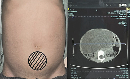
Figure 1. Anterior view of the patient’s abdomen
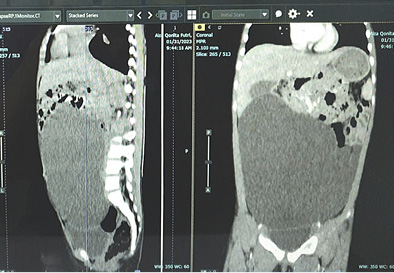
Figure 2. Computed tomography of the abdomen demonstrated a AP 7.10 x LL 15.64 xx CC 14.97 cm cystic mass
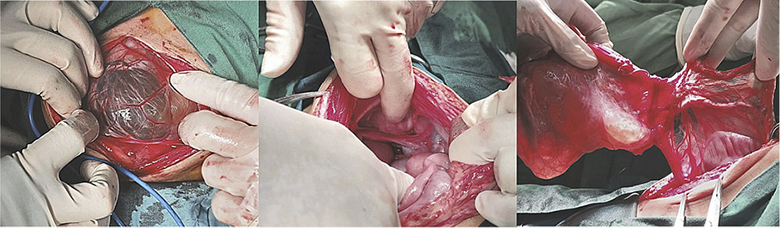
Figure 3. Intraoperative finding showing omental cyst, the origin, and the cyst wall
It was determined by anamnesis, physical examination, and imaging that the diagnosis was an ovarian cyst. As we continue laparotomy with midline incision from obstetrics department, we revealed a 8x8x10 cm sized cystic mass within the left transverse colon omentum, uterus and both ovaries were normal (Figure 3). We excised the cyst in toto and omentectomy then sent for pathological anatomy and cytology analysis. Instead of an ovarian cyst, we found an omental cyst during the operation (Figure 4), thus we had to conduct an excision and omentectomy and postoperatively achieved favorable results. Pathological examination revealed findings a inclusion cyst containing connective tissue, fat, and muscle. Cytology findings are suggestive for transudative effusion from the cyst and the international system for reporting serous fluid (ascites) cytopathology negative for malignancy (Figure 5). She was discharged three day after.
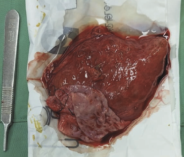
Figure 4. Cut-open omental cyst
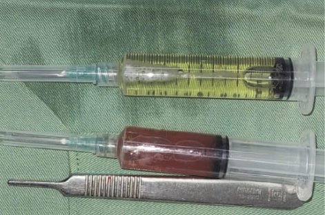
Figure 5. Serous fluid aspirated from cyst and serosanguinous fluid from intra-peritoneal
Discussion
Omental cysts are rare intra-abdominal tumors in children, which are reported incidence of one in twenty thousand admissions to a children’s hospital and female predominance by a factor of 1.5. Different etiological factors have been reported for development of these masses including a benign proliferation of mesenteric lymphatics, failed fusion of the mesenteric leaves, and deficiency of the lymphatico-venous shunts. The literature contains several of cysts which are definitely congenital, but there are also acquired cysts which have been reported. A female factor were found in our case. Presentation is often with abdominal pain with distention and/or palpable abdominal mass, a very small cyst may be asymptomatic and nonpalpable. Disorders of the respiratory or urinary system, in addition to symptoms caused by compression of the portal vein, are often seen because of cyst enlargement. In our case we found a abdominal distension with palpable mass for two months with no history of abdominal pain, and no disorders of respiratory or urinary system. These cysts are usually diagnosed incidentally by abdominal imaging or discovered as an incidental finding at operation. Since it is a rare condition and there are lack of specific symptoms, a correct pre-operative diagnosis is difficult. Our patient underwent CT scanning, allowing us to locate the cyst precisely and identify its relation to the surrounding tissues, leading to a diagnosis of ovarian cyst at first. In laparotomy, we found a giant omental cyst, not originating from the ovaries, and did an omentectomy to excision the cyst. CT scan shows that the giant abdominal cyst is difficult to identify between omental, mesenteric, and ovarian cysts. Precise identification of stomach cysts is necessary. Also, doctors need to read the CT scan image more carefully.
Conclusion
Giant omentum cysts are difficult to identify clinically and imaging from ovarian and mesenteric cysts. There is a need for a more detailed history, physical examination, and support, as well as a more thorough reading of the CT scan.
References
1. Alemu H, Alemu S, Berhane M. Omental Cyst Presenting as an Acute Abdomen in a Pediatric Patient: A Case Report. Int Med Case Rep J 2022; 15: 43–46.
2. Robbins KJ, Antiel RM, Shakhsheer BA. Omental Cyst: A Case Report and Review of the Literature. Ann Pediatr Surg 2021; 17(1): 62.
3. Sayeed M, Benzamin M, Akter S, Mazumder MW, Karim AB, Dey BP. Omental Cyst – Rare Cause of Abdominal Pain in a 7-Year-Old Child: A Case Report. GE Port J Gastroenterol 2021; 28(3): 202–206.
4. Bassiouny IE, Abbas TO. Omental Cysts in Children: Rare Causes of Abdominal Masses (A Report of Two Cases). Open Journal of Pediatrics 2013; 3(4): 418–421.
5. Alemu H, Alemu S, Berhane M. Omental Cyst Presenting as an Acute Abdomen in a Pediatric Patient: A Case Report. Int Med Case Rep J 2022; 15: 43–46.
6. Kao NH. A Case Report for a Diagnostic Dilemma of a Giant Intra-Abdominal Cyst with an Uncertain Origin. Int J Surg Case Rep 2020; 71: 374–377.