Lietuvos chirurgija ISSN 1392–0995 eISSN 1648–9942
2024, vol. 23(1), pp. 52–55 DOI: https://doi.org/10.15388/LietChirur.2024.23(1).7
Post Varicella Necrotising Fasciitis Afflicting Upper Limb in Young Child: A Rare Case Report
Swarup Prabhu
Command Hospital Air Force Bangalore, Bengaluru, India
E-mail: swarupprb@gmail.com
Ayjaz Hussain
Command Hospital Air Force Bangalore, Bengaluru, India
E-mail: drayjazhussain@gmail.com
Tarun Gupta
Command Hospital Air Force Bangalore, Bengaluru, India
E-mail: tmgupta26@gmail.com
Abstract. Necrotising fasciitis is a common surgical emergency. Risk factors include immunosuppression, type 2 diabetes mellitus, chronic diseases like cirrhosis of liver and chronic kidney disease, malignancies, intravenous drug use, obesity, and age more than 60 years. Our case is of a 14 years old boy who presented with necrotising fasciitis (NF) of right upper limb post varicella of 10 days duration. Prompt diagnosis with aggressive surgical debridement was lifesaving in this case. Early reconstruction with split skin grafting escalated recovery resulting in a fully functional limb without any residual deficit. This case is a rare example of post varicella NF in a young child without any co-morbidities. Majority such cases are encountered by a general surgeon in an emergency setting. Hence, this report sensitises and enlightens the first responders about the existence of this rare entity.
Keywords: necrotising fasciitis, varicella, immunosuppression, debridement.
Received: 2023/10/11. Accepted: 2023/11/12.
Copyright © 2024 Swarup Prabhu, Ayjaz Hussain, Tarun Gupta. Published by Vilnius University Press. This is an Open Access article distributed under the terms of the Creative Commons Attribution Licence, which permits unrestricted use, distribution, and reproduction in any medium, provided the original author and source are credited.
Introduction
Chickenpox is a highly infectious disease of childhood caused by varicella zoster. Two percent of children with varicella develop complications, of which bacterial superinfection of skin is the most common complication (45%) [1]. Necrotising fasciitis (NF) is a rare but potentially lethal complication of varicella, seen in <1% of the children, who develop bacterial skin superinfection [2].
Case report
14-year-old boy, a diagnosed case of varicella for past 10 days, on home-based management presented to our hospital with painful swelling of right forearm with overlying blisters and foul smelling discharge associated with high grade fever of 4 days duration. He had no known co morbidities, was developmentally normal, adequately nourished and immunised for his age.
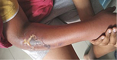
On general examination, the child was irritable and sick looking. He was febrile with 102.2 ºF, had tachycardia and tachypnoea, with signs of early shock. Pleomorphic varicella lesions were noted all over the body. Local examination revealed a tender, warm, oedematous right forearm with few blisters and necrotic patch of skin measuring 4×3 cm in size associated with purulent, foul-smelling discharge on the lateral aspect of forearm (Figure 1). Right elbow had painful restriction of movements while shoulder and wrist joint had a full range of movements. There was no distal neurovascular deficit and both right radial and ulnar artery were palpable. Systemic examination was unremarkable. Calculated Laboratory Risk Indicator of Necrotising Fasciitis Score (LRINEC) score was 11 (Table 1) which was strongly predictive of NF.
Table 1. Lab investigations with Laboratory Risk Indicator of Necrotising Fasciitis (LRINEC)
|
Lab test |
Values |
LRINEC Score |
|
Haemoglobin |
8.2 |
2 |
|
Total count |
28 100 |
2 |
|
Differential LC (N/L*) |
N 35/L 50 |
|
|
Platelets |
141 000 |
|
|
RBS |
193 mg/dl |
1 |
|
Sodium/Potassium |
125/3.4 |
2 |
|
Blood urea |
10 |
|
|
Creatine |
0.3 |
|
|
ESR |
54 |
|
|
CRP |
227 |
4 |
|
Total LRINEC score |
|
11 |
*N – neutrophil, L – lymphocyte, LRINEC – Laboratory Risk Indicator of Necrotising Fasciitis, ESR – erythrocyte sedimentation rate, RBS – random blood sugar.
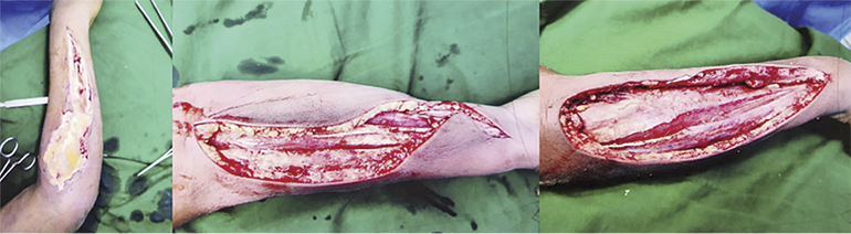
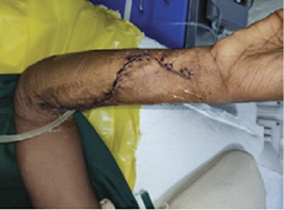
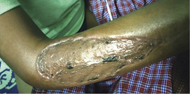
He underwent emergency debridement and fasciotomy of flexor and extensor compartment of right forearm under general anaesthesia with axillary block (Figure 2) on day of presentation. Intra operatively dish water pus, necrotic skin and subcutaneous tissue, oedematous muscles of extensor group of right forearm were noted. Drained pus and tissue were sent for culture and sensitivity, which showed growth of methicillin-resistant Staphylococcus aureus (MRSA). Blood culture showed no growth. Post operatively he was managed with culture specific antibiotics and daily normal saline dressings for 4 days. On post operative day (POD) 5 secondary suturing was done for flexor compartment fasciotomy wound and two sessions of negative-pressure wound therapy (NPWT) of 5 days each were given for post debridement wound over extensor compartment of right forearm. Split thickness skin graft (SSG) was harvested from left thigh and placed over the wound on POD 15 (Figure 3). Wound review done on day 5 post SSG revealed graft uptake of 100%. Physiotherapy sessions were given for right elbow till 2 weeks post operatively. On follow up visit after 6 weeks both donor and graft sites had healed well. The operated limb was fully functional with no restriction of joint movement at elbow or wrist.
Lab investigations with Laboratory Risk Indicator of Necrotising Fasciitis (LRINEC) score were as follows:
|
Figure 1. Necrotic patch right forearm |
|
|
Figure 2. Post debridement and fasciotomy (intra-operative) (intra-operative images showing dish water pus and post debridement and fasciotomy wound) |
|
|
Figure 3. Secondary suturing for fasciotomy wound of flexor compartment on POD 5 |
Figure 4. Post-operative day 10 following SSG for wound on extensor compartment with 100% graft uptake |
Discussion
Chickenpox is a common contagious infection of childhood. It usually resolves without complications. Bacterial skin superinfection is one of the most common complications of chickenpox [3].
Necrotizing fasciitis is an aggressive soft tissue infection that tracks along the skin and subcutaneous tissue involving the superficial fascia [4]. The overall incidence of NF due to various aetiologies in children is 0.08/100 000 children per year [5].
Bacterial superinfection of the varicella skin lesions is quite common, but progression to necrotizing fasciitis is seen in only 1% of these cases [2]. Necrotizing fasciitis is of four types: Type I (polymicrobial), Type II (monomicrobial), Type III (marine organisms (Vibrio vulnificus)), Type IV (fungal species) [6]. Type II infections are less common, caused by invasive Group A beta haemolytic streptococci and sometimes by staphylococci. They typically occur in healthy patients with no significant past medical history [7]. 70% of post-varicella NF is caused by group A beta haemolytic streptococci and is associated with 13.6% mortality [5]. Immunocompetent children who develop varicella usually get type 2 NF [8].
Initially, patient may present with mild pain and swelling of involved area and can be misdiagnosed as cellulitis. In NF there is severe pain disproportional to the extent of lesion associated with paraesthesia. Skin changes in form of bullae, crepitus, necrotic patches and foul-smelling discharge appear later in the course. Patient may rapidly progress to frank sepsis and multi-organ dysfunction syndrome (MODS) without aggressive surgical management [9].
In our patient, we enlisted NF, haemorrhagic varicella and purpura fulminans as the differential diagnosis. As the child was developmentally normal and previously immunocompetent, haemorrhagic varicella was ruled out. In purpura fulminans, there should have been presence of palpable purpuric lesions becoming confluent, and leading to well-demarcated eschar with features of disseminated intravascular coagulation [10]. Nonsteroidal anti-inflammatory drugs (NSAIDs) use is associated with an increased incidence of NF [8]. However, our patient gave negative history for the same.
At admission, a maximum of 30% of cases are diagnosed accurately as NF. There is 100% mortality if surgical debridement is not done [11]. NF involving the limbs have a better prognosis than those involving head and trunk [9].
Conclusion
NF requires high index of suspicion in every case of post-varicella cellulitis. It requires early and aggressive surgical debridement, vigorous fluid resuscitation and appropriate antibiotic cover to prevent life and limb loss [12].
Declarations
Financial Disclosure. Nil.
Ethical approval. Patient consent and ethical approval taken from institutional ethical committee.
Conflict of Interest. All authors declare no conflict of interest.
References
1. Clark P, Davidson D, Letts M, Lawton L, Jawadi A. Necrotizing fasciitis secondary to chickenpox infection in children. Can J Surg 2003; 46(1): 9–14.
2. Nirmala C, Madhusudan K, Venkateswara RJ, Satish K. Necrotising fasciitis in an infant secondary to varicella zoster infection. J Dr NTR Univ Health Sci 2013; 2: 55–57.
3. Kapse A. Chicken pox: Though seldom it could be a menacing malady even in healthy children. Paediatr Infect Dis 2009; 1: 154–160.
4. Stevens DL, Bisno AL, Chambers HF, Dellinger EP, Goldstein EJC, Gorbach SL, Hirschmann JV, Kaplan SL, Montoya JG, Wade JC. Practice guidelines for the diagnosis and management of skin and soft tissue infections: 2014 update by the Infectious Diseases Society of America. Clin Infect Dis 2014; 59(2): e10–e52.
5. Bingöl-Koloğlu M, Yildiz RV, Alper B, Yağmurlu A, Ciftçi E, Gökçora IH, Ince E, Emiroğlu M, Dindar H. Necrotizing fasciitis in children: diagnostic and therapeutic aspects. J Pediatr Surg 2007; 42(11): 1892–1897. DOI: 10.1016/j.jpedsurg.2007.07.018.
6. Giuliano A, Lewis F Jr, Hadley K, Blaisdell FW. Bacteriology of necrotizing fasciitis. Am J Surg 1977; 134(1): 52–57. DOI: 10.1016/0002-9610(77)90283-5.
7. Anaya DA, Dellinger EP. Necrotizing soft-tissue infection: diagnosis and management. Clin Infect Dis 2007; 44(5): 705–710. DOI: 10.1086/511638.
8. Xavier R, Abraham B, Cherian VJ, Joseph JI. Early diagnosis of post-varicella necrotising fasciitis: A medical and surgical emergency. Afr J Paediatr Surg 2016; 13(1): 44–46. DOI: 10.4103/0189-6725.181707.
9. Sadasivan J, Maroju NK, Balasubramaniam A. Necrotizing fasciitis. Indian J Plast Surg 2013; 46(3): 472–478.
10. Chalmers E, Cooper P, Forman K, Grimley C, Khair K, Minford A, Morgan M, Mumford AD. Purpura fulminans: recognition, diagnosis and management. Arch Dis Child 2011; 96(11): 1066–1071.
11. Puvanendran R, Huey JC, Pasupathy S. Necrotizing fasciitis. Can Fam Physician 2009; 55(10): 981–987.
12. Claudia CS, Margareta IM, Sorina CD, Ionescu C. Necrotizing fasciitis with group A Streptococci and Eggerthella lenta as a complication of Varicella in a child – case presentation. ARS Med Tomitana 2014; 20(1): 35–39.